Adipose tissue: structure and functions
Adipose tissue is a collection of body cells that primarily serve to store energy in the form of fat. Adipose tissue is also responsible for thermal insulation of the body and mechanical protection of organs (coating them with a fat pad). In addition, adipose tissue also performs an endocrine function: it releases some necessary substances into the blood.
Adipose tissue is divided into two types: white and brown. The first type can be either white or yellowish; the second species has a characteristic brownish-brown color. This color of the fat layer occurs due to the presence of a large amount of cytochrome, an iron-containing pigment.
Brown adipose tissue warms the human body because it produces heat. An adult has a small amount of brown adipose tissue, which is located near the kidneys and thyroid gland; babies have much more of it, and it disappears as they grow older.
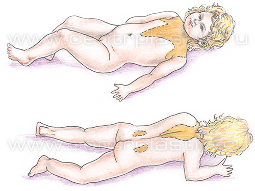
Distribution of brown adipose tissue in a newborn
Distribution of brown adipose tissue in the adult human body
In addition to white and brown, there is so-called mixed adipose tissue, consisting of the two types listed above. It is located between the shoulder blades, on the chest and shoulders of a person.
The fat cell is designated by the term "adipocyte". This name is of mixed Greek and Latin origin: the Latin element "adeps" means "fat", the Greek word "kytos" means "hollow bubble".
A scanning electron microscope allows you to look at the cells of adipose tissue and see that they look like balls surrounded by collagen fibers and capillaries with blood.
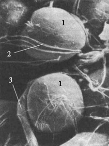
Photograph of adipose tissue cells.
1 - Adipose tissue cells; 2 - Collagen fibers; 3 - Capillary
Most of the fat cell is a voluminous bubble of fat enclosed in a membrane; the cell nucleus and mitochondria are pushed to the periphery, and the nucleus takes on a flattened shape.
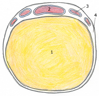
Adipose tissue cell.
1 - Fat vesicle; 2 - Cell nucleus; 3 - Mitochondria; 4 - Cell membrane
Adipose tissue is formed during the development of the embryo from connective tissue - mesenchyme, which is the basis for all types of connective tissues in the body.
This happens as follows: a mesenchymal cell is transformed into a lipoblast, and this, in turn, becomes a mature fat cell - an adipocyte.
An interesting fact is that humans are one of the few mammals that are born with ready-made fat deposits, which are formed after 30 weeks from the beginning of intrauterine development.
Previously, doctors believed that the number of ready fat cells does not change in a person throughout life. Now this point of view is considered erroneous, since although mature cells do not divide, cells that are the precursors of fat cells, which are precisely capable of dividing, are preserved.
There are two periods in a person’s life during which fat progenitor cells actively multiply and thereby increase the number of adipocytes:
- embryonic development
- puberty.
As a rule, in other periods, precursor cells do not multiply, and further weight gain is possible only by increasing the size of those fat cells that already exist. This change in adipose tissue is called hypertrophic growth.
For comparison: 35 billion and 125 billion fat cells
But no cell can increase in size indefinitely. Therefore, if the amount of fat in a cell approaches a critical limit, a signal is given to progenitor cells, which trigger the proliferation mechanism, creating new fat cells. Their number can increase significantly: for example, a thin adult has approximately 35 billion fat cells; the number of them in someone who suffers from severe obesity can reach 125 billion.
This change in adipose tissue is called hyperplastic (hypercellular) and can occur at any age.
If new fat cells have already formed, then with weight loss they do not disappear, but only decrease in size.
Most fat deposits are found under the skin and in the abdominal area. The fat layer in those who are overweight can reach a thickness of 15-20 cm.
These layers are not homogeneous, they are “slices” measuring 5-10 mm.
Adipose tissue is divided into two layers: superficial and deep. In turn, these layers consist of three layers of adipose tissue, called apical, mantle and deep.
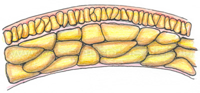
The uppermost, apical layer of tissue is adjacent to the skin and serves as a kind of “cover” for sweat glands, hair follicles and blood vessels. The next layer, the mantle layer, consisting of fatty pearls, is located in the middle and makes up the most voluminous part of the adipose tissue. The thinnest layer is the deep one, which covers muscle tissue.
The fat cells of the body are characterized by a strict sequence and hierarchical structure. The layer of adipose tissue consists of segments formed from “pearls”, which in turn are formed from lobules - groups of lipocytes (fat cells).
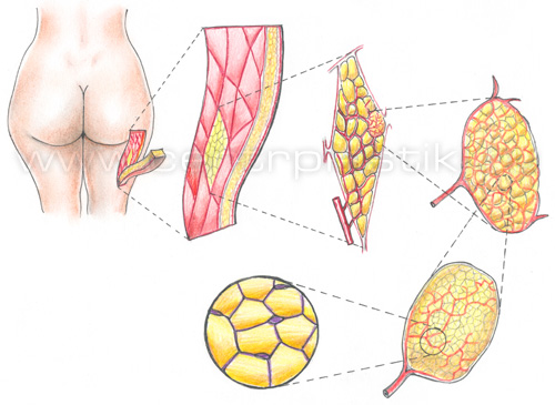
Fat deposition in the abdominal area can occur not only in the subcutaneous space, but also in a special abdominal organ called the omentum. The fat cells in this organ can collect and retain significant amounts of fat.
Also, large fat deposits are found in the retroperitoneum, the place where important organs are located: kidneys, pancreas, aorta, etc.
Fat deposits distributed unevenly in our body.
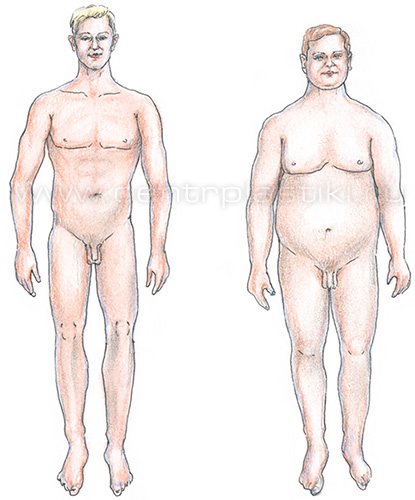
Excess weight is characterized by two types of fat deposition: central and peripheral. Depending on the type of deposits, in popular literature, such types of figures as “apple” and “pear” are sometimes distinguished.
The central type of obesity is characterized by the formation of fat deposits mainly in the abdominal cavity (which is why it is called abdominal).
Peripheral obesity is accompanied by the deposition of fat mostly under the skin.
As it turned out as a result of research, these two types of fat deposits differ in their roles. The central type of obesity is accompanied by the deposition of metabolically active brown fat around the internal organs. Peripheral obesity provokes the deposition of metabolically inactive white fat.
The main functions of fat in the body
Energy storage
Fat occupies 65-85% of the total weight of the adipocyte (fat cell), presented in the form of triglycerides (also called triacylglycerols). Their main function in the body is breakdown, releasing large amounts of energy. Overweight people have a huge amount of energy in reserve in the form of triglycerides. It would be enough to provide basic metabolism for several months.
Fats are the most “beneficial” substance for storing energy. Per unit weight, fats contain twice as much energy as carbohydrates, since they can be present in the body in pure form and in large quantities.
One kilogram of fat is calculated to contain energy equal to 8750 kilocalories.
Thermal insulation
Some animals store fat under the skin for two purposes at once: firstly, it serves as a thermal insulation layer that protects the body during cold weather, and secondly, fat serves as an “energy depot”. Thick layers of triglycerides are a distinctive feature of seals, walruses, penguins and other warm-blooded animals of the Arctic and Antarctic.
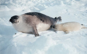
Harp seal. The very thick layer of subcutaneous fat of this animal serves not only as a fat depot, but also plays the role of a reliable warm “wetsuit.”
Mechanical protection
Fatty tissues of the body not only protect internal organs from mechanical damage, but also control their location in the body. For example, it is known that the kidney has a “fat cushion” that holds it in place, so kidney prolapse only threatens very thin people.
The fatty tissue present around the eyeball also holds it in place and protects it from direct contact between the eye and the orbital bones.
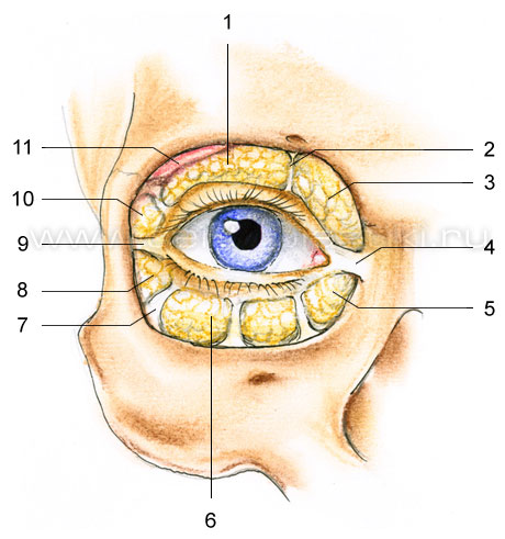
1 - Intraorbital fat - central portion; 2 - Dividing partition; 3 - Intraorbital fat - internal portion; 4 - Internal canthus; 5 - Intraorbital fat - internal portion; 6 - Intraorbital fat - central portion; 7 - Ligaments; 8 - Intraorbital fat - outer portion; 9 - Outer canthus; 10 - Intraorbital fat - outer portion; 11 - Lacrimal gland
Endocrine function
Modern research suggests that adipose tissue is not just a place where energy reserves are stored. They are actively involved in the production of hormones, i.e. can be classified as endocrine organs. Two hormones that are secreted by fat cells have already been thoroughly studied - leptin and estrogens.
Leptin was first isolated in 1994 and has been hailed as a potential anti-obesity drug. As doctors assumed, when leptin is released by fat cells, it enters the brain, causing a feeling of fullness. But, as further experiments showed, administering leptin to a person during meals did not provoke a feeling of satiety.
As it turned out later, leptin is a regulator responsible for the time that passes between meals. Thus, the higher the leptin level, the less often a person eats. But since overweight people have more leptin in their blood than they should, its use as a medicine does not make sense.
Estrogens. Adipose tissue has aromatase activity because it contains the aromatase P450 enzyme, which converts testosterone, the male sex hormone, into female sex hormones called estrogens. The rate of conversion increases with age, as well as with the growth of fat accumulation.

Fat cells take up testosterone from the blood and release estrogens into it. Fat accumulated in the abdomen has particular aromatase activity. Thus, it becomes clear why in men, when a “beer belly” appears, almost “female” breasts appear, and why obesity leads to a decrease in potency and fertility.